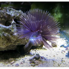
Latin Polychaeta, from Greek πολύς - many, Greek χαίτη - hair.
Polychaeta - class Annelida. Length from 2 mm to 3 m. The body consists of several hundred segments with parapodia (skin growths) and bundles of bristles located on them. On the head section are located sensory organs. Predominantly bottom-dwelling, free-living. Over 7000 species. Are of great importance in the diet of fish and other aquatic animals.
Polychaeta are characterized by an average size of about 10 cm in length and 0.5–1.0 cm in width. Interstitial species are usually small, their size does not exceed 1 mm. On the other hand, among the Polychaeta there are large species reaching a meter in length. The largest Eunice aphroditois is 3 m long. The body consists of many (sometimes up to several hundred) rings-segments, in each of which a complex of internal organs is repeated: paired coelomic sacs, associated genital ducts and excretory organs. A distinctive feature is parapodia — lobe-shaped appendages extending from each segment of the body, bearing chitinous bristles (chetes). In some species, the function of the gills is performed by the corolla of the tentacles on the head area. There are eyes, sometimes complicated, and balance organs (statocysts).
The vast majority of representatives are inhabitants of marine waters. They inhabit the ocean from the tidal strip (littoral) to the Abyssal. Adults, as a rule, are bottom forms. The greatest depth at which Polychaeta are found is from 10,160 to 10,730 m. In the World Ocean, Polychaeta inhabited almost all bottom biotopes and pelagials — from interstitial to hydrothermal oases of oceanic rift zones. Very few Polychaeta live in fresh water (for example, the genus Manayunkia in Lake Baikal), in the forest floor and in the soil at a depth of more than 3 m (genus Parergodrilus and genus Hrabiella). Most lead a burrowing lifestyle. The next place in number is occupied by crawling Polychaeta. Many species can swim, and some even lead a pelagic lifestyle all the time. Finally, there are Polychaeta, which form tubes and lead a sedentary lifestyle in them. Parasites are very rare among Polychaeta, but some species parasitize other Polychaeta, starfish and fish. The stage of life of larvae of some species from the families Amphinomidae, Sabellariidae, Oweniidae, Magelonidae is crossing the Atlantic Ocean currents from Africa to America. They are called transoceanic larvae. Many of them manage to swim across the ocean in 2-3 months, then settle to the bottom, turn into adult worms and live in the coastal area.
The prostomium (head blade) is the very first segment (the count of body segments begins with the peristomium), always one, although it can be segmented a second time. In practice, when determining the boundaries of the prostomium, it is convenient to use the position of the mouth opening: the prostomium is a segment located above or in front of the mouth, often forming the upper lip. The prostomium can carry appendages. If these appendages are approximately the same size and shape, then they are called antennae (head tentacles). If one pair stands out sharply among the appendages, then this pair is called palps (tentacles). In some taxa, the palps may merge (Syllidae), or split into two pairs (some Onuphidae), or form a gill-trapping apparatus (Serpulomorpha), or oral tentacles (Terebellomorpha), or shift to the peristome (Spionidae). Both antennae and palps can have supports, called, respectively, cerato- and palpophores, their distal parts are called cerato- and palpostyles. In addition, there can be variously arranged eyes on the prostomium: from simple pigment spots to complex optical systems. The eyes can be located both directly on the surface of the prostomium, and on special outgrowths — ommatophores. The form of the prostomium often has taxonomic significance.
Among the trunk segments, the peristomial segment (segments) and the body segments themselves are distinguished. The peristomial segment — the peristomium — follows directly after the prostomium, with the peristomium the counting of body segments begins. It carries a mouth opening from the abdominal side, but the following segments (in Amphinomidae and Spintheridae) can also take part in the formation of the mouth opening. The peristomium, as well as the segments following it, may differ sharply from those located behind them. Usually, in the latter case, the segments following the peristomyium are very similar to it. In this case, they are called peristomial segments. The peristomium may partially or completely merge with the prostomium and/or the following segments. Often this merging is so complete that in some cases there is still no consensus about the initial number of segments and their boundaries. Fused peristomium and peristomial segments are often referred to as peristomium for short. Often the peristomium bears peristomial (tentacular) antennae similar in structure to the antennae, but unlike them, the peristomial antennae are homologous to the appendages of the parapodia. The number of pairs of peristomial antennae approximately corresponds to the number of segments that formed the peristomium (1-2 pairs per segment). The oral opening located on the peristomium usually leads to a more or less everting or protruding pharynx. The pharynx in many families is armed with chitinous jaws (which can calcify), and its surface can be covered with soft leathery papillae and/or chitinous teeth (paragnates, chevrons, etc.).
The pygidium (pygidium) is usually cone-shaped with an apically or slightly dorsally displaced anal opening located at the posterior end. As a rule, the pygidium bears tendril—like sensitive appendages - urites. Their number and size vary widely. Often it is a pair of well-developed antennas. Some (Protodrilus, etc.) may develop suction devices on the pygidia (papillae, blades, etc.). There are often eyes on the Sabellidae pygidia.
Body segments, as a rule, carry parapodia on the sides — more or less pronounced outgrowths of the body, equipped with bristles. The boundaries between segments are often poorly defined. There can be only one pair of parapodia on each segment. Each parapodia can have one or two branches equipped with bristles: dorsal — notopodia and abdominal — neuropody. Parapodia are used by worms when crawling along the bottom of the reservoir, and when the animal swims, they play the role of fins. In worms that dig in the ground or live in tubular houses, parapodia are completely or partially reduced.
The integuments of Polychaeta consist of a single-layer cuticular epithelium and are almost completely devoid of atrial fibrillation cells. The latter are preserved only in larvae, where they form shimmering hoops covering the head blade and then each metamer. Ciliary locomotion exists at this stage of ontogenesis before the transition to movement with the help of parapodia. The ciliary method of locomotion and, accordingly, the ciliary hoops are preserved in adult individuals of those Polychaeta that are considered to be secondarily crushed forms of neotenic origin (Ophryotrocha). Finally, the ciliated epithelium is preserved in some forms not for locomotion, but for creating water currents through the body, necessary under certain living conditions. In particular, this is why the ciliated cells are preserved on the gills of a number of Polychaeta. Fields of scintillating cells are also found in short-lived Polychaeta, in which they can, for example, carry food particles to the mouth opening. Or, on the contrary, remove feces from the tube. The cuticle of Polychaeta is dense and flexible at the same time, not having such a powerful, complex layered structure as in nematodes. Its structure varies in different Polychaeta, representing either a thick single layer of polysaccharides and albumin proteins, or a lattice of collagen fibrils alternating with granular layers. In this case, ultramicroscopic studies reveal two layers in the cuticle: the epi- and basal cuticle. The first consists of a fine-grained or fine-fiber matrix and an electron-dense layer in which peculiar spherical bodies are located. Acidic mucopolysaccharides have been found here. The basal cuticle consists of fibrils of polysaccharide or glycoprotein nature, forming a powerful disordered or, conversely, an ordered lattice of collagen fibers. In all cases, the cuticle is not solid. Epidermal cells form numerous ultramicroscopic protrusions — microvilli, which penetrate through the pores of the lattice of fibrils and partially even rise above the surface of the cuticle. Thanks to this, there is a free exchange of substances between the epidermis and the environment. The epidermis of Polychaeta contains a large number of glandular cells. They can open on the surface of the cuticle. Among the glandular cells, mucosal cells predominate, which produce mucus to protect the body (for example, in Anaitides mucosa). The epithelium is underlain by a layer of tissue of the main substance in the form of a thin border plate with highly developed precollagen fibers and usually devoid of cells. The covers of worms leading an active life at the bottom of the reservoir are distinguished by a well-developed cuticle. On the contrary, worms swimming in the water column, burrowing into the ground or building tubular houses have a very thin cuticle. The secretions of glandular cells serve as a building and cementing material in the construction of tubes in which some polychaete worms live.
The musculature of the Polychaeta lies directly under the border plate. It is represented by a layer of annular muscles located on the outside and, deeper, a layer of longitudinal muscles. There is also a dorsoventral musculature, which in Polychaeta is closely associated with the development of the walls of dissepiments and mesenteric coeloma. There are also muscle bundles related to the system of locomotor organs (parapodia) and special muscle formations of various organs (intestines, blood vessels, etc.).
The digestive tract of Polychaeta consists of three divisions: anterior ectodermal (stomodeal) — pharyngeal; middle (endodermal) — assimilating; posterior (proctodeal) — excretory. The stomodeal section begins with a transverse oral opening, followed by the oral cavity, muscular pharynx (pharynx) and esophagus ("esophagus"). Many predatory worms have chitinous teeth in the throat, which serve to grasp prey. Polychaeta demonstrate a great variety in the structure of the stomodeal department. This is especially true of farinx. This is due to the fact that they have mastered a wide variety of biotopes and very different strategies for obtaining food. The stomach is available only in sedentary Polychaeta. Nereis have large blind processes of the esophagus that secrete digestive enzymes. The endodermal midgut, unlike the anterior part of the intestine, which is quite mobile along the longitudinal axis of the body, is firmly at rest, fixed by dissepiments and covered along its entire length by mesenteria. It is usually straight, without twists, which is a consequence of its fixation by the walls of the coelomic sacs. The transport of food particles in the stationary intestine is carried out mainly with the help of cilia. To do this, as a rule, along the ventral surface of the midgut there is a longitudinal groove or roller bearing ciliated cells. In the middle intestine, food is digested and nutrients are absorbed. The short ectodermal posterior intestine opens terminally with the anal opening. However, the powder may shift dorsally or ventrally. For species living in tubes, different forms of excrement removal are characteristic, ensuring the withdrawal of these structures from the tube to the outside. The most original method is characteristic of representatives of the Sabellariid family. These species live in vertically arranged tubes, and their front end faces upwards. The posterior part of the body of these Polychaeta is greatly reduced, only the fecal excreting tube lying in the back of the dorsal side of the animal remains. From the end of this tube along the dorsal side of the body to the anterior end (up) stretches the ciliary groove, along which dense feces are removed from the tube.
Respiration in small forms is carried out by the surface of the epidermis. In larger species, areas are formed in certain places of the epidermis, equipped with a network of vessels and a powerful capillary network associated with it. Some species have special thin-walled outgrowths of the body or parapodia, which are specialized respiratory organs, such formations are traditionally called gills. In crawling forms, the dorsal antennae of the parapodia most often turn into gills, if any. The need for gills arose as a result of a strong decrease in the amount of oxygen diffusing through the integuments dissolved in water, which is a consequence of a reduction in the relative surface of the body due to an increase in its linear dimensions and the development of cuticular integuments. The shape of the gills is highly variable: there are filamentous (in the simplest case), bubble-like, comb- and tree-like. In a number of Polychaeta, spurs of coelomic sacs enter them, in others, in addition, the gills are equipped with a network of capillaries of the circulatory system.
The transport functions of Polychaeta are carried out by the circulation of fluid in the whole and in the circulatory system. If the dissepiments are completely lost (which is typical for representatives of the Glyceridae family), then the circulatory system is also lost, transport functions are then carried out only by the whole. In some small Polychaeta, the circulatory system may also be absent. The structure of the circulatory system of Polychaeta corresponds to the general scheme of the circulatory system of annelids. It is closed, includes two main longitudinal vessels — the pulsating dorsal and non-contracting abdominal. Metameric vessels are represented only by transverse vessels that do not make up a complete ring (as in the case of annular metameric vessels), they are directed from the dorsal and abdominal main vessels to the body wall, parapodia or any other organs, where they are divided into a system of capillaries (after the capillaries are combined into larger vessels, the blood returns through them to which-or from longitudinal vessels). Two transverse vessels depart from the abdominal vessel, one of which carries blood to the dorsal branch of the parapodia, the other to the abdominal one. The same vessels supply blood to the body wall and gills, if any. Blood from the parapodia and the body wall enters the spinal vessel. From there, the blood goes to the intestinal plexus, from the intestinal plexus to the abdominal vessel. Respiratory pigments can be represented by hemoglobin, which is the most common variant, hemerythrin and chlorocruorin. Hemoglobin and chlorocruorin are in the blood plasma, hemerythrin, the presence of which is characteristic of one family of Polychaeta, is contained in blood cells.
As a general rule, there is a pair of protonephridiae or a pair of metanephridiae in each torso segment of the Polychaeta. Nephridia are characterized by ectodermal origin. The inner end of each nephridium lies in the whole, near its back wall. The nafridium canal penetrates the dissepiment and in the next segment opens outward with a hole — a nephropore — on the side of the body. Protonephridia are characteristic of small Polychaeta species that do not have a circulatory system. Large Polychaeta species tend to have metanephridia. The latter are convoluted tubes that begin in the body cavity with a funnel with cilia. The outer end of the metanephridium opens either directly outward or into the longitudinal common excretory canal. The final part of the metanephridium has an expansion — the bladder.
The nervous system is represented by the supra-pharyngeal ganglion (brain) and the abdominal nervous ladder or abdominal nervous chain (abdominal brain). In forms with well-developed sensory organs, the supra-pharyngeal ganglion can acquire large dimensions and be divided into several lobes. The brain plays the role of an independent center that regulates the activity of the entire nervous system of the trunk. As the norm, it is located in the head blade. Dorsally and laterally, it is usually not separated from the integumentary epithelium, whereas from below it is underlain by a subepithelial border plate. The volume of the brain varies greatly: in errant large Aphroditidae, the total number of brain cells reaches 14 thousand cells, and in Polydora ciliata — sedentary form — barely reaches 800. In its most developed form, the abdominal brain is a pair of powerful nerve trunks running ventrally along the entire length of the body parallel to each other. As a rule, they are brought together along the medial line up to the point of merging into a single anatomical whole, but in Serpuloidea, for example, they are quite noticeably removed from each other and, thanks to the commissures (transverse nerves), form an abdominal nervous ladder. The depth of the abdominal brain also varies markedly. In some Polychaeta, it lies subepithelially (Phyllodocidae, Nephtydidae, etc.). All stages of immersion of the abdominal brain from under the cover into the body wall, under the annular musculature and further into the coeloma cavity are traced in a number of families of Polychaeta. Typical for Polychaeta is the presence of pedal ganglia located at the base of the parapodia. Tentacles and palps carry mechanoreceptors (tactile) and chemoreceptors on their surface. Peristomial antennae, like tentacles and palps, are equipped with mechanoreceptors and chemoreceptors. In the back of the prostomium there are photosensitive organs. These organs are represented in different numbers and can be simple eyes consisting of only two cells, or rather complex eyes, including thousands of sensing cells, as well as lenses. More complicated eyes are found in swimming species. In addition to the prostomium, the eyes can be located on body segments, on the pygidium, on the gills. On the prostomium there are sniffing organs, or olfactory pits — small depressions of the integument, at the bottom of which there are ciliated cells that perform a chemoreceptor function.
Most Polychaeta are segregated. Each segment contains a pair of gonads. They lie in connective tissue and are associated with septa, blood vessels and the coelomic lining. Gonads are often present in all segments, but in some Polychaeta they are confined to special genital segments. Hermaphrodite Polychaeta are few in number. Among them there are species (some Sabellidae) in which the anterior abdominal segments produce eggs, and the posterior ones produce sperm. As a rule, gametes are excreted into the external environment through metanephridia or through ruptures of the body wall.
The nutritional characteristics of different Polychaeta are closely related to their lifestyle. Many burrowing forms and sedentary inhabitants of burrows and tubes are ground—eaters using organic matter contained in the soil. Some of them swallow the soil directly with their mouth or with the help of a pharynx equipped with a bulbus, which is able to bulge outwards and is characterized by weak muscle development. These are the so-called swallowing detritophages, which include, for example, Arenicolidae, Maldanidae and Polychaeta earthworms from the groups Orbiniidae and Capitellidae. In contrast, collecting detritophages, such as representatives of Terebellidae and Spionidae, first collect organic particles with specialized appendages, which then send food into the mouth. Representatives of Terebellidae collect food with the help of prostomial tentacles capable of stretching and contracting, and Spionidae do this with long peristomial palps. Carnivorous and herbivorous forms and Polychaeta scavengers are usually represented by mobile (wandering, or errant) forms that crawl on the substrate or catch Alciopidae, but some of them live in tubes or actively dig in the ground. To capture prey or carrion, these Polychaeta have a well-developed, muscular, ever-turning throat. The inverted pharynx may have the shape of a muscular tube and serves to capture prey (Phyllodocidae). Sometimes it is equipped with a thick, grater-like lower lip (Amphinomidae) or bears jaws with prehensile (Nereidae, Eunicidae, Lumbrineridae) or poisonous (Glyceridae) teeth. The horny jaws are built of tanned protein. As soon as the food is detected, the pharynx is instantly turned out. At the same time, the jaws located at its end are brought forward and open. The jaws grab the food, after which the pharynx is retracted just as quickly. Although in some cases Polychaeta have special protractor muscles, the eversion of the pharynx, as a rule, occurs due to an increase in the pressure of the coelomic fluid as a result of contraction of the muscles of the body wall. When these muscles relax and the pressure drops, the pharynx is retracted by the retractor muscles. All Polychaeta that feed on suspension (sestonophages) are sedentary animals living in tubes. The latter can be submerged in the ground or attached to clam shells, rocks and other hard surfaces. To capture particles suspended in water, such Polychaeta use special appendages bearing cilia and having a large surface area (in Sabellidae, for example, it is a feathery "crown") or secrete a mucous network through which water is actively driven and filtered (Chaetopterus). A very small number of Polychaeta species lead a parasitic lifestyle. Labrorostratus (Oenonidae) settles in a whole other Polychaeta and can reach almost the same size as the host. Some misostomids are parasitic in starfish. Ectoparasites include the blood-feeding Ichthyotomidae, which attach to the fins of some marine fish.
Most Polychaeta easily restores lost body parts. In general, the ability to regenerate is better expressed in homonomous forms. Experimental studies have shown that in some species it is possible to completely restore the body from just one segment. Regeneration is under the control of the nervous and endocrine systems.
Polychaeta implement various ways of movement. Burrowing Polychaeta move in the ground due to peristalsis. In some burrowing Polychaeta, the pharynx takes part in the movement, which, turning forward, anchors in the ground, then the worm contracts the pharynx and pulls forward. Sometimes this kind of movement is carried out only due to the work of the pharynx. When crawling, the parapodia and bristles work like legs, alternately pushing off from the substrate during the working stroke and rising above the substrate during the return movement. The movements of numerous parapodia are well coordinated. Parapodia located on opposite sides of one segment move in opposite phase. The movements of the parapodia located on one side of the body are shifted relative to each other by a fraction of a phase. The undulation of the body in the lateral direction also takes part in the crawling movement. Similar movements are used by polychaetes when swimming.
Fertilization in Polychaeta is external. They lay eggs directly into the water, where their fertilization takes place and pelagic larvae (trochophores) develop from them, more or less existing in the water column for a long time and capable of active nutrition. Some of the Polychaeta species living in the ground or in the interstitial have mastered internal fertilization and, accordingly, have copulatory organs. There are species in which sperm is injected into the female's body through the skin, due to its piercing, in other cases the spermatophore is glued with the help of a copulatory organ to the surface of the female's body, the spermatozoa drill through the skin with their own means and enter the female as a whole. In some species with internal fertilization, there is concern for the offspring, expressed in the formation of a protective cocoon for laying and even wearing it on the female's body. After a certain period of planktonic life, the trochophora sinks to the bottom, where metamorphosis occurs. The anterior hemisphere of the trochophora forms tentacles and palps, as well as other organs, and gradually turns into a prostomium. The perioral section of the trochophora is rebuilt into a peristomium. The posterior hemisphere of the larva is strongly elongated and is divided into several segments at once, which are called larval segments. The posterior end of the posterior hemisphere is transformed into a pygidium. A pair of coelomic sacs develops in each segment. In the process of matamorphosis, the brain is formed, abdominal nerve trunks are laid on the abdominal side in the form of a paired ectoderm roller. In the future, they come into contact with the brain with the help of near-pharyngeal connectives. From the ectoderm, sensory organs develop — eyes, palps. Thus, the next larval stage, the metatrochophore, is formed from an unsegmented, primary—cavity trochophore, characterized by segmentation and a metameric coelome. The metatrochophora swims for a while or leads a bottom lifestyle without changing significantly, and then experiences further metamorphosis. At the leading edge of the anal lobe, a growth zone is formed, the cells of which are continuously multiplying. The area lying in front of it consists of rapidly growing yet undifferentiated tissues. In this zone, new segments (postlarval) are formed and successively one after another are separated in the direction of the front. The process continues until as many segments are formed as there are in an adult worm. Consequently, the body of an adult animal consists of departments of different origin: the head blade or prostomium, which is a modified anterior hemisphere of the trochophora; several larval segments; numerous postlarval segments and anal lobe or pygidium originating from the posterior part of the trochophora. The musculature of the musculoskeletal sac and intestines, the lining of the coeloma, gonads and coelomoducts are formed from the mesoderm, the nervous system, metanephridium channels, anterior and posterior intestines are formed from the ectoderm, the middle intestine is formed from the endoderm. Some species are able to reproduce asexually. There are two variants of asexual reproduction: archetomy and paratomy. In the case of an archetomy, the worm's body is first divided into fragments, and then completes the front and rear ends of the body. Paratomy implies the reverse sequence of events: during this process, a chain of worms linked to each other by different ends of the body is formed. Sexual dimorphism, as a rule, is not pronounced. The most well—known cases of such are the epitoxic stages of Polychaeta from the families Nereidae and Syllidae. Sexual dimorphism occurs only in mature individuals preparing for spawning. In this case, the anterior (atopic) part of the body of the polychaete, devoid of gonads, differs both in the structure of the parapodia, which, as a rule, are less developed, and in body coloration. The latter is due to the color of sexual products that shine through the translucent integuments of the body. This leads to the appearance of sexual dimorphism, since the sexual products of males and females are colored differently. The posterior part of the body, in addition to coloration, is characterized by a greater thickness, more developed parapodia and is called epitoxic.
Polychaeta
Tags: polychaeta