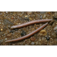
Lancetnidae (Branchiostoma) - a genus of small to 8 cm long marine animals with lanceolate translucent body, belong to the Lancetniformes (Branchiostomatiformes), class Lancetnidae (Leptocardii), subtype Craneless. For Lanceletniki characterized by - full segmentation of the body, the absence of the skull, brain and highly developed sensory organs, the presence of atrial (perigastric) cavity, neural tube and chorda, which stretched from the front to the rear end of the body. Marine. Dwell in warm and temperate waters, mainly at depths of 10 to 30 meters.
The structure of lancelets represents the basic scheme of the structure of all chordates. It retains all the features of chordates throughout its life. The general plan of their structure includes all the characteristic features of this type:
the presence of chordae;
gill slits that penetrate the pharynx;
the nervous system in the form of a neural tube; the chorda is between the neural tube and the intestine;
ventral position of the anus and the presence of a tail, which does not include the intestine, but does include other axial organs - the chorda and the neural tube.
The body of lanceolates is translucent, whitish to creamy-yellow, sometimes with a tinge of pink, with a faint metallic luster, compressed from the sides and elongated. It is pointed at the posterior end and obliquely cut at the anterior end, the ventral side is slightly wider than the dorsal side. The length of the body of lanceolates varies between 5-8 cm. On the underside of the anterior end of the animal is a preoral funnel (or cavity) surrounded by oral tentacles. Along the entire back stretches a fin fold - a low dorsal fin. It is transparent and supported by numerous rod-like fin rays. The dorsal fin passes without a visible boundary into the caudal fin, which is lance-shaped or lanceolate. The caudal fin functions as an engine. On the ventral side, along the lower edge of the tail, there is a short sub-caudal fin (also erroneously called pelvic fin). The boundary of the caudal and sub-caudal fins is marked by the anal opening. All these fins, oriented in a plane of bilateral symmetry, perform the function of stabilizers during movement, that is, do not allow the animal to turn over. From the front end of the body to the caudal fin on the sides go so-called metapleural folds. At the point of convergence of the metapleural folds and the caudal fin is located atriopore, or gill pore, - the outlet of the atrial, or perigastric, cavity.
Lancet skin is a single-layer epithelium (epidermis), which is located on the underlying thin basal membrane. From above, the epidermis is covered with cuticle, a superficial film of mucopolysaccharides secreted from epidermal glands, which protects the thin skin of lancelets from damage. Beneath the epithelium is a thin layer of gelatinous connective tissue - corium, or cutis. The outer covers are transparent, almost not pigmented.
The central nervous system of lanceolates is a neural tube lying above the chorda with a narrow cavity inside - the neurocelia. The anterior end of the neural tube is shorter than the chorda; this feature gave the subtype the name "cephalochordates". The brain and spinal cord are not externally differentiated, but the head and spinal parts of the neural tube have a different structure and perform different functions. The right and left sides of each segment of the neural tube are connected by nerve cells forming reflex arcs and neurons.
The sensory organs are primitive. Tactile sensations are perceived by nerve endings of the entire epidermis, especially the oral tentacles. Chemical stimuli are perceived by encapsulated nerve cells, which are also located in the skin and lining the fossa Kelliker. In the neural tube, mainly in the area of its cavity, there are photosensitive cells with concave pigment cells - Hesse's eyes. Translucent animal covers freely pass light rays, which capture the eyes Hesse. They also work as photorelays, registering the position of the lanceolate's body in the substrate.
The axial skeleton of lanceolates is the chorda, or notochord. It is a light-colored, vertically striated rod that extends, thinning, along the dorsal side of the body from the anterior end to the posterior end. In lanceolates, the chord extends far forward into the cephalic end, behind the neural tube. The notochord of lancelets is a unique formation, which has no analogues among other representatives of the type of chordates. The chorda is released from the entoderm during neurulation, lacing off the dorsal side of the primary intestine, which borders the myogenic (muscle-forming) complex. The notochord consists of a complex system of transverse vacuolized epithelial-muscular plates and is surrounded by a sheath of gelatinous connective tissue. The plates are far apart from each other and only in some places are connected by thin transverse outgrowths. Notochord functions like a muscular organ: muscle contraction causes an increase in its rigidity. The axial skeleton of lancelets has the properties of a hydrostatic skeleton. The lanceolate chord, together with the neural tube, is surrounded by a connective tissue sheath with which the septa between myomeres - myosepta - are connected.
The lanceolate musculature is metameric, that is, it has a segmental structure and extends on both sides of the chorda. Myomeres (or myotomes) adjacent to the chorda are muscle segments (50-80 in number), separated by myosepts - jelly-like connective tissue septa. Myosepts are fused with the chorda sheath and cutis, a thin layer of connective tissue of the skin. Each myomer is shaped like half a cone, the apex of which is inserted into the notch of the next segment toward the anterior end of the body. This is how the connection between the myomeres and the axial skeleton is maintained. Asymmetry of musculature is characteristic for lanceolates - each muscle segment on one side of the chorda is displaced by half in relation to the myomeres of the other; the location of the myoseptum is opposite to the middle of the myomeres of the opposite side. Sometimes this type of segmentation, also characteristic of many representatives of the Vendian fauna, is called sliding reflection symmetry. From the head end of the body to the atriopore, a special layer of unsegmented transverse muscles runs along the belly of the lanceolate. Lancelets move worm-like, due to the contraction of myomeres, consistently bending the body (undulatory movement). Bending, elastic tail blades push the body of the lanceolate forward. The lanceolate is able to burrow into the ground with its tail forward.
On the lower part of the head end there are oral tentacles and a preoral funnel leading to a small mouth opening. It is surrounded by a muscular annular membrane, the sail. The sail acts as a partition between the mouth opening and the vast pharynx. The front part of the sail is covered with thin ribbon-like outgrowths of the mesenteric organ, the rear part has short tentacles directed into the pharyngeal cavity; they are an obstacle to large food particles. The pharynx in lanceolates occupies up to one third of the body length and is penetrated by gill slits numbering over 100 pairs. Gill slits are separated by inter-gill septa with ciliated epithelium and lead into the pharyngeal cavity, or atrial cavity, rather than directly outside (thus, the gill slits are not visible from the outside; they are covered with protective skin folds). The atrial cavity surrounds the pharynx on the sides and bottom and has an opening to the outside, the atriopore. In the form of a blind closed growth, the atrial cavity extends slightly beyond the atriopore. The movements of the growths of the mesenteric organ and the oscillations of the cilia covering the inter-gill septa, direct the slow and non-stop flow of water into the pharynx. Then the water passes through the gill slits into the perigastric cavity, and from there it is discharged through the atriopore. The pharynx has two furrows lined with ciliary and glandular epithelium. The gill furrow (gill groove, endostyle) runs along the lower part of the pharynx, and the supra gill furrow (supra gill groove) runs along the dorsal side of the pharynx. They are connected by two bands of ciliary epithelium that run along the lateral inner surfaces of the pharynx in its anterior part. The cells of the endostyle secrete mucus, which under the action of the flickering of cilia is driven to the anterior end of the pharynx - towards the flow of water. Along the way, food caught in the pharynx is enveloped and captured. After that, mucus glued lumps of food on the two semicircular grooves move into the suprazhaber furrow, through which they race back to the initial part of the intestine (intestine). The mucus, in which the food was wrapped, flows down the sides of the pharynx and forms a mucous membrane on the gill slits, allowing water to pass outside. Narrowing sharply, the pharynx passes into a short, unbent intestine, which ends in the anus. At the point of transition of the pharynx to the intestine is located blind finger-shaped hepatic outgrowth, which secretes digestive enzymes. It is located on the right side of the pharynx and is directed toward the head end of the lanceolate. Digestion takes place in the cavity of the hepatic outgrowth and in the entire intestine.
The excretory system of lancelets is compared to the nephridial system of ringed and flatworms. It is something between the protonephridial and metanephridial systems. About 100 pairs of nephridia are metamerically located above the pharyngeal cavity. They have the appearance of a short, steeply curved tube opening with an orifice into the atrial cavity. Almost all of the rest of the nephridia enter as a whole (supraglottic ducts). This part of the tube has nephrostomies - few openings closed by a group of solenocytes, specialized cells with a "flicker flame" - a constantly working flagellum. To the walls of the nephridium tube adjoin capillary tubules, through the walls of which metabolic products enter the integument. From the integument, decomposition products penetrate into the solenocyte, and from there - in the lumen of the nephridial tube, through which they move with the help of beating flagella solenocytes and cells of the mesenteric epithelium lining the tube. From there, through the opening of the nephridial tube, the wastes enter the perigastric cavity and are excreted from the body of the lancelet. In addition to the nephridia located in each metamere, the lanceolate has an unpaired (left) nephridium of Gatchek, which is the first to appear in ontogeny. In its structure, it resembles the other nephridia. For decades, the origin of the lanceolate's protonephridia remained unclear. Old authors (Goodrich et al.) were inclined to the opinion about their ectodermal origin (for example, Goodrich described their development from unicellular rudiments, which, in his opinion, belonged to the ectomesoderm). Thus, it was assumed that the lanceolate nephridia were not homologous to the mesodermal nephrons (kidneys) of vertebrates. Recently, molecular biological data supporting the mesodermal origin of the lanceolate nephridia have been accumulating.
The respiratory system is characterized by the fact that there are no specialized organs. Gas exchange is carried out through the entire body surface. The importance of the circulatory system for gas exchange is also controversial due to a number of reasons: lanceolates have no heart (pulsating vessels contract uncoordinatedly), no endothelium, erythrocytes and respiratory pigments. Therefore, diffusion plays an important role in the process of gas exchange in lanceolates. The coelomic cavities of the lanceolate are quite extensive, and their walls contain contracting myoepithelial cells. In addition, the muscles and coelomic cavities are adjacent to the inner and outer surfaces of the body of the lanceolate - the epithelial layer of the atrial cavity and the skin. This arrangement is ideal for direct gas exchange. Thus, it is quite possible that the main circulatory system transporting oxygen and carbon dioxide is the coelomic system.
The circulatory system is partially closed and separated from the surrounding organs by the walls of blood vessels. Small vessels are devoid of endothelial lining, the endothelium of large vessels is not continuous. Beneath the pharynx is the abdominal aorta (aorta ventralis), a large vessel whose walls are constantly pulsating and pumping blood, thus replacing the heart. Pulsation occurs through slow, uncoordinated contraction of the myoepithelial layer of the adjacent coelomic cavities. Through the abdominal aorta, venous blood travels to the head end of the body. Through the thin cover of hundreds of gill arteries (outflow arteries), branching by the number of gill septa from the abdominal aorta, the blood absorbs dissolved oxygen in water. The bases of the gill arteries - bulbs - also have the ability to pulsate. The gill arteries flow into the paired (right and left) roots of the dorsal aorta (aorta dorsalis), which is located at the posterior edge of the pharynx, and extends under the chorda to the end of the tail. The anterior end of the body is supplied with blood by two short branches of the paired roots of the dorsal aorta (aorta dorsalis) - the carotid arteries. The branching arteries of the dorsal aorta supply blood to all parts of the body. This is how the arterial circulatory system of lancelets is represented. After passing through the capillary system, from the walls of the intestine venous blood is collected in the unpaired axillary vein, going as a hepatic vein to the hepatic outgrowth. In it, the blood is again scattered into capillaries - the portal system of the liver is formed. The capillaries of the hepatic outgrowth again merge into a short hepatic vein, flowing into a small extension - the venous sinus. From both ends of the body, blood collects in the paired anterior and posterior cardinal veins. On each side they merge to form the right and left cuvier ducts (common cardinal veins), flowing into the venous sinus, which is the beginning of the abdominal aorta. From this it follows that lancelets have a single circulatory circle. Their blood is colorless and does not contain respiratory pigments. Oxygen saturation of blood in arteries and veins is similar - small size of animals and single-layer skin allow oxygenation of blood not so much through gill arteries, but through superficial vessels of the body.
They live in many temperate seas, including the Black Sea, in coastal areas with clean sandy bottoms. As lanceolates are benthic animals, they spend most of their time on the bottom, taking different feeding positions depending on the looseness of the sand. If the sand is loose, lancelets burrow deeply into the substrate and expose only the front end of the body; if the soil is silty and dense, the animals lie on the bottom. When disturbed, lancelets can swim a short distance and burrow again or lie on the ground. They can also move through wet sand. Lancelets live in colonies of more than nine thousand individuals per square meter. They make seasonal migrations - swim several kilometers. Lancetniki - animals-filterers: food is absorbed through the mouth with the flow of water, which is driven by the movement of cilia. Food for lancelets is mainly phytoplankton and plankton - various branchiopod crustaceans, infusoria, diatom algae; as well as larvae and eggs of other lower chordates and invertebrates. The nature of the diet is thus passive. Oxygen, as well as food, enters the body of the lanceolate with the flow of water.
Lancelets reproduce in spring, summer or fall. Immediately after sunset, females begin to throw mature eggs (eggs). Fertilization occurs in water, as does the subsequent individual development of lancelets. Embryonic development of lanceolates is used in many textbooks and manuals as an example to describe the embryogenesis of chordates, as it represents a simplified scheme of development of all higher chordates.
Lancetes
Tags: lancetes


