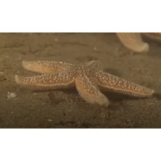
Echinodermata is a type of marine invertebrates. Body length from a few millimeters to 5 m (fossils up to 20 m). Echinodermata have radial, usually five-beam symmetry. Calcareous skeleton formed by numerous needles, spines and other appendages. Body shape is stellate, globular, disc-shaped, etc. Echinodermata have a unique, absent in all other animals, ambulacral (water-vascular) system, which serves for movement, excretion and touch. They are widely distributed, living in all seas and oceans. Reproduction is most often sexual. Development with free-swimming - larva and subsequent meta-morphosis. Some Echinodermata (sea urchins, holothurians) are fishery objects.
There are about 7000 modern species. Along with chordates and semichordates, the Echinodermata belong to the branch of the secondary horned animals (Deuterostomia). Modern representatives of the type are sea stars, sea urchins, ophiurae (snaketails), holothuria (sea cucumbers), and sea lilies. Some representatives, such as holothurians, are used for food. The type also includes approximately 13,000 extinct species that have flourished in the seas since the early Cambrian.
Adult Echinodermata are characterized by radial and usually five—ray symmetry of the body, while their larvae are bilaterally symmetrical. Thus, Echinodermata possess a secondarily acquired radial symmetry of the body. All Echinodermata undergo a five-ray stage of development, even if they eventually regain bilateral symmetry (sea cucumbers, irregular sea urchins). Many sea lilies and some starfish have a large number of arms, usually multiples of five. In some Gorgonocephalus, the arms branch out, forming a complex tree-like structure. In adult Echinodermata, there is a distinction between the oral side, on which the mouth is located, and the opposite aboral side, on which the anal opening is usually located. The oral side of actively moving starfish, ophiura and sea urchins faces the substrate on which the animal crawls. The body of sea cucumbers is elongated in the oral-aboral direction: at one end of it is the mouth, and at the other — the anal opening. Many stalked sea lilies lead a sedentary lifestyle, attaching to the substrate with a stalk that departs from the aboral side of the calyx. On the oral side of the rays (arms), ambulacral legs are usually located, with the help of which starfish and sea urchins move. The planes of symmetry of Echinodermata, in which rows of ambulacral legs are located, are called radii. Interradiuses are located between the radii. The external radial symmetry of the animal is violated by a madrepore plate located on one of the interradiuses. The size of Echinodermata varies from a few millimeters to 2-3 meters (large holothurias), and in some extinct species — even up to 20 m (stalked sea lilies with long stems). The body of starfish and ophiurians has a pentagonal or star—shaped shape, sea urchins have a spherical, heart-shaped (Echinocardium cordatum) or disc-shaped (flat sea urchins) shape, in holothurians the body is barrel-shaped or worm-shaped, and in sea lilies it resembles a flower.
In the subcutaneous layer of Echinodermata, a biomineral endoskeleton develops, which consists of calcareous plates and often forms a variety of external appendages: needles, spikes and pedicellariae. In many sea urchins, these appendages develop especially strongly. Needles perform a protective function. They are often mobile. Some sea urchins point their needles towards the approaching danger. Pedicellariae are needles modified into grasping tweezers. With their help, the animal is cleaned and gets rid of parasites. In sea urchins, the skeleton takes part in the formation of a special chewing organ — the Aristotelian lantern. In ophiura, the skeleton is especially developed in the rays, where it forms a series of massive calcareous segments — vertebrae. In holothurians, the skeleton is reduced. Echinodermata, unlike all other animals, can reversibly change the stiffness of their integuments and connective tissue. They have connective tissue capable of changing its rigidity — the so-called mutable connective tissue. Extreme stiffness values vary: when a starfish bends in an arc over a victim (for example, a mollusk), it stiffens its connective tissue and its rays become a support for ambulacral legs that attach to the flaps of the mollusk. After eating, the connective tissue softens, becomes elastic, and the starfish straightens. Sea urchins, by changing the stiffness of connective tissue, can fix the position of needles, which are used to repel predators or to anchor in rock crevices. Under stressful conditions, ophiura and holothuria spontaneously reject (autotomize) rays or eject internal organs with the help of local softening of connective tissue. In extreme cases, when some holothurias are removed from the water into the air, their body completely softens, spreads, and the animal dies. Although the integuments of Echinodermata contain muscles, nerves and other types of cells, it is the extracellular matrix of connective tissue that changes stiffness. There are nerve cell endings in this matrix, and there are probably two types of nerves: the action of some makes the matrix rigid, the action of others softens it. The stiffness of the matrix is affected by changes in the concentration of Ca2+ and other cations. In general, an increase in the concentration of Ca2+ gives the matrix rigidity, and a decrease softens it. Ca2+ can participate in the formation of bridges between macromolecules in the matrix. The epidermis of Echinodermata includes mechanoreceptor cells that provide touch, pigment cells that cause the color of the animal, and glandular cells that secrete mucus or toxins.
The mouth of Echinodermata is located on the oral side of the body. The anal opening is most often located on the aboral side, but in sea lilies and irregular sea urchins it is located on the oral side of the body. Ophiurs do not have an anus. Most often the digestive tube is represented by a long intestine. In sea stars, a voluminous stomach is formed, capable of turning inside out through the mouth. Starfish envelopes the stomach of the prey, which can not swallow, and thus performs external digestion. The digestive glands are represented by hepatic outgrowths and rectal glands.
Some Echinodermata have an organ (functionally integrated heart, axial canal, and axial blood vessel) that is homologous to the reno-pericardium ("heart-kidney") of semichordates, but its role in excretion is questionable. Other Echinodermata have no such organ, and all have excretion at the cellular level.
One of the most original features of the structure of Echinodermata should be considered the complex differentiation of the integument into a number of systems: body cavity, ambulacral (water-vascular) and perigemal systems. The internal organs of Echinodermata lie in the extensive body cavity. The body cavity wall consists of peritoneal epithelium, a single layer of squamous cells that surrounds all the viscera. Some organs lie in pockets of the body cavity and as if suspended on special folds - mesenteries. In the skin of Echinodermata there are outgrowths, which enter the body cavity - skin gills, which perform a respiratory function. Their wall is very thin, so through it easily occurs gas exchange. Holothuria form special respiratory organs - water lungs. The body cavity is filled with coelomic fluid containing numerous amoeboid cells. These cells absorb waste products and foreign bodies and leave the body through the covers. Thus, they perform excretory and immune functions.
The ambulacral system is unique for Echinodermata. It is a network of canals communicating with the environment through the stony canal and the madrepore plate, filled with a fluid with a composition close to sea water. From the radial ambulacral canals branch many ambulacral legs, at the base of which are ampullae - muscle vesicles, when contracting which leg lengthens. At the end of the leg is a suction cup. The ambulacral system is involved in respiration, locomotion and food extraction. For example, with the help of the cooperation of many ambulacral legs, a starfish can open the shell of a bivalve mollusk.
The perigemal system is a set of channels and cavities (sinuses) surrounding the animal's circulatory system. The circulatory system is poorly developed and is a system of cavities in connective tissue (lacunae) without endothelial lining. Each ray contains two radial perihemal canals, with a radial blood vessel in the septum between them. The radial vessels flow into the oral blood ring lying in the septum between the two annular perihemal canals. The genital sinus surrounds the aboral blood ring and the genital stolon. The two blood rings are connected by an axial organ surrounded by the left and right axial sinuses.
An axial organ complex is located in one of the interradii of Echinodermata. It includes organs from different systems:
A stony canal that connects the annulus ambulacralis to the madreporal plate;
The axial organ, within which a network of blood vessels is located;
Left axial sinus - The part of the cecum that connects the inner annular perihemal canal to the right axial sinus;
The right axial sinus, which is capable of rhythmic contraction and thus facilitates the movement of blood in the vessels, i.e., it performs the functions of the heart;
Sexual sinus - a section of the coelom containing the sex cord, which consists of immature sex cells.
The nervous system of Echinodermata is primitive, consisting of three separate parts built on a radial plan: the nerve ring and radial nerve tracts. In the oral body wall lie two nervous systems - sensitive and motor. In the aboral body wall - only motor. Sensory organs of Echinodermata are quite diverse, but primitive in structure. They are diffusely distributed throughout the body in the form of various sensitive cells (functions of touch, chemical sense, vision). Light-sensitive cells can be assembled in the oculars. In starfish, the eyes are located at the ends of the rays, and in sea urchins around the anus.
Most Echinodermata are bisexual animals, form many small, yolk—poor eggs and sweep them into the water. Fertilization in Echinodermata is external. The development of the embryo occurs in water, as part of the meroplankton. The fertilized egg (zygote) begins to break up and after a while forms a blastula. Crushing is complete, radial type. Usually Echinodermata hatch from an egg at the blastula stage. Such a blastula-like larva is a bubble, the wall of which consists of one epithelized layer of flagellate cells (blastomeres), and the cavity of which (blastocele) is filled with gelatinous fluid. The blastula is able to move with the help of flagella. Often, at the animalic pole of the larva, the flagella are longer and perform a sensory function (apical organ). After some time at the vegetative pole, some cells are evicted (immigration) into the blastocele. Cells of the primary mesenchyme involved in the formation of the larval skeleton. After that, the vegetative wall is implanted (invagination) into the blastocele, resulting in the formation of a closed primary intestine (archenteron). At the same time, cells of the secondary mesenchyme are evicted from the apex of the archenteron into the blastocele, which have amoeboid mobility and contribute to invagination, and later to the isolation of coelomes, the formation of the mouth and larval arms. Then the separation of the coelomic mesoderm, which is part of the archenteron, occurs by forming a bulging wall, which is unlaced in the form of a coelomic sac, that is, by the enterocellular method. In the future, the whole is divided first into right and left, and then into three pairs of coelomes: right and left axoceles (anterior), hydroceles (middle) and somatoceles (posterior). Usually the left coelomes are ahead of the right ones in development, which is due to their leading role in metamorphosis. This completes the gastrulation process, as a result of which three germ leaves are isolated: ectoderm (larval integument), endoderm (gut) and mesoderm (primary and secondary mesenchyme, as a whole). The blastopore shifts to the abdominal side and becomes a powder (anus). At the anterior end, on the abdominal side, an ectoderm is formed, the so-called oral bay (stomodeum), which merges with the intestine and forms the mouth. The intestine is divided into three sections: the esophagus, the enlarged stomach and the small intestine. The body acquires an ovoid shape. A perioral cavity forms around the mouth, along the edge of which a ciliary cord is formed, on the rest of the body surface the cilia disappear. With the help of the beats of the cilia of the cells of the ciliary cord, the larva moves and adjusts the food to the mouth. This stage of bilaterally symmetrical larva, common to most Echinodermata, is called dipleurula. In the future, the larva acquires adaptations to the planktonic lifestyle, different in different classes, and becomes pluteus (sea urchins and ophiura), auricularia (holothuria) or bipinnaria, and later — brachiolaria (starfish). In species with a large amount of yolk, lecithotrophic (that is, yolk-feeding) barrel-shaped doliolaria larvae, girdled with transverse ciliated rings, develop from the egg. After some time, free-swimming bilaterally symmetrical larvae begin metamorphosis, as a result of which they transform into a radially symmetrical adult animal. In the body of the larva, the rudiment of the future adult animal (imaginal disk) is formed. On the left side of the larva, the oral side of the animal is formed, and on the right — the aboral side. By the end of metamorphosis, there is a complete reduction of larval organs.
Along with sexual reproduction in Echinodermata there is in some cases and sexless reproduction, when the body of the adult organism is divided in half or into several pieces. Each half regenerates the lost parts. In a certain connection with sexless reproduction is very characteristic for Echinodermata ability to restore lost individual organs or parts of the body - regeneration. Very often under unfavorable conditions or when attacked by an enemy Echinodermata discard (autotomize) rays or parts of the body, throw out viscera, and sometimes even disintegrate into pieces. The rate of recovery of lost parts is different and very dependent on temperature.
They live almost always on the seabed from the littoral and practically to the extreme depths. At greater depths, Echinodermata, mainly holothurians, are the dominant group of bottom animals. They do not tolerate changes in water salinity, as they are unable to regulate the salt composition of body fluids. Many Echinodermata are detritophagous, there are polyphagous (many ophiura), predatory (most starfish) and herbivorous (many sea urchins). Most Echinodermata have the ability to regenerate (repair) body parts. For example, an entire animal can regenerate from a single sea star ray.
Thanks to the mineralized skeleton, Echinodermata are well preserved in the fossil state (although they are often represented only by scattered skeletal elements), and the peculiar structure of these elements (stereome) makes it possible to easily identify them. The oldest known fossils of Echinodermata date back to the third century of the Cambrian (about 515-520 million years ago), when they appear approximately simultaneously in the seas of Laurentia, Angara and Gondwana. These are representatives of helicoplacoidea, edrioasteroidea and eocrinoidea. Hypotheses about belonging to Echinodermata of some Precambrian organisms (for example, Arkarua) have not received widespread support. Representatives of the five modern classes are known since the Lower — Middle Ordovician. The heyday of the type falls on the Paleozoic. The closest relatives of Echinodermata among modern animals are the semichordates, forming together with them the Ambulacraria clade. This clade is a sister group of chordates. The time of separation of the semichordates and Echinodermata by the molecular clock method is estimated at about 580-550 million years ago. The common ancestor of all the secondborns was a bilaterally symmetrical free-living animal with three pairs of coelomic sacs. This is indicated by the presence of a stage of development similar in all secondary-mouthed. In Echinodermata, this stage corresponds to a dipleurula larva. The appearance of the first Echinodermata is associated with the transition of this hypothetical ancestor to a sedentary lifestyle and its acquisition of radial symmetry. In the fifth century of the Cambrian, representatives of the Carpoidea class appeared, which existed until the Lower Devonian. They led a sedentary lifestyle, but did not yet possess radial symmetry. The body was covered with plates, the mouth and anus were placed on the side facing away from the substrate. The internal organs were located asymmetrically. Representatives of the Cystoidea class have radial ambulacral grooves around the mouth, designed to collect food from the water column. The rest of the Pelmatozoa originate from the globules: the Blastoidea class, modern sea lilies and the Edrioasteroidea class, which included free-living species. The first Eleutherozoa, combining the features of modern starfish, ophiura and sea urchins, belonged to the class Ophiocistia. From them, the modern representatives of the subtype originated.
Echinodermata
Tags: echinodermata